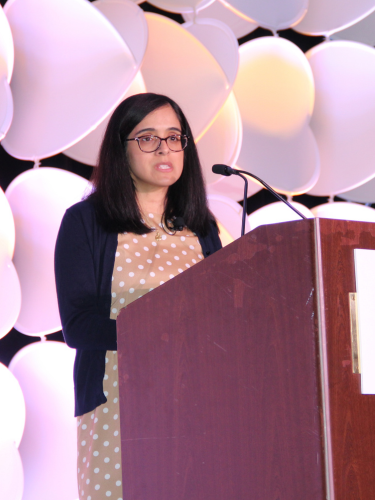
Interesting Cases in Pediatric Dermatology
Pediatric dermatologist, Dr. Garza-Mayers highlighted interesting cases she’s managed in a fascinating presentation at the SDPA conference in Boston. The first case presentation revealed an infant with scalp and facial lesions which worsened with sun exposure. This patient was ultimately diagnosed with neonatal lupus erythematosus (LE). Up to half of these patients will present with skin lesions, with “raccoon eyes,” a pathognomonic finding. This condition is transferred passively maternally. It is important to know that most mothers will have asymptomatic connective tissue disease. This condition is associated with congenital heart block; therefore, it is essential these patients are referred to pediatric cardiologists for evaluation. Patients with neonatal LE must be counseled to practice sun protection. Skin lesions will typically clear by a year of age. There is a risk this will recur in up to 25% of subsequent pregnancies.
Coverage: SDPA Annual Summer Dermatology Conference, June 22-25, 2023 – BOSTON
Annular erythema of infancy presents with enlarging erythematous annular patches and plaques and is a benign diagnosis of exclusion. These patients do not present with systemic findings and the condition tends to resolve without residual scarring or atrophy within a year. The common presentation of infantile hemangiomas was explored by Dr. Garza-Mayers. While most hemangiomas are benign, it is important to evaluate these patients for the PHACES acronym, (posterior fossa anomalies, hemangioma, arterial anomalies, cardiac anomalies, and eye anomalies) to determine the need for further work-up. Diagnosis of infantile hemangiomas is typically clinical and doesn’t require a biopsy. If patients present with > 5 hemangioma lesions, evaluation with abdominal ultrasound is recommended to help identify any internal hemangiomas of the organs. Treatment of hemangiomas include topical timolol, oral propranolol and laser treatment.
Dr. Garza-Mayers reviewed the differential diagnosis of café au lait lesions. It is typically considered normal to have up to 5 café au lait lesions, therefore, patients who present with > 6 café au lait lesion need to be evaluated for neurofibromatosis 1 (NF1). The rate of NF1 is one in 3000 births with 50% occurring due to a spontaneous mutation. Additional findings that may correlate with NF1 include nevus anemicus and juvenile xanthogranulomas. Lastly, juvenile localized scleroderma conditions were explored including morphea en coup de sabre. Patients who present with localized scleroderma need to have a full skin examination including a genital exam to check for lichen simplex et atrophicus. Localized scleroderma is not on the spectrum of systemic sclerosis.
By Sarah B.W. Patton
